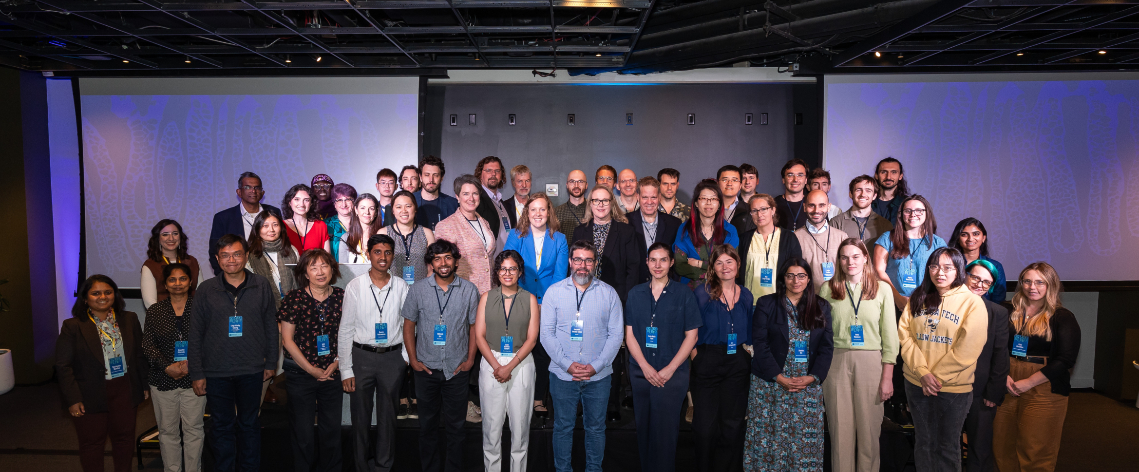
For the past 6 years, the Helmsley Charitable Trust has convened researchers of the Gut Cell Atlas Crohn’s Disease Consortium (GCA) to share their progress in cataloging cell types and their signals in the intestine of Crohn’s disease (CD) patients. By generating this searchable cellular atlas of diseased tissue – and comparing it to that of healthy tissue – researchers hope to uncover key mechanisms of disease initiation and progression towards the development of new therapies and personalized care.

Dr. Alexandra-Chloe Villani presenting at GCA 2024.
To create an atlas, researchers isolate single cells and analyze their gene expression profile. These cells are then assigned an identity (annotation), and similar cells from datasets across research studies are combined into a common atlas (integration). Building a universal gut cell atlas for the community presents unique challenges. Cells can take on new identities or aberrant functions in a diseased environment, obscuring cell annotation. Differences in sample processing and disease phenotypes also complicate the integration of data across institutions. Dr. José Ordovas-Montañes and Dr. Alexandra-Chloe Villani discussed how these challenges are being met both in the GCA and the global Human Cell Atlas.
With atlasing underway, new biology is emerging that could shift our approach to treating disease by filling key knowledge gaps.
An intact intestinal barrier, made of tightly connected epithelial cells, is a fundamental feature of a healthy intestine and separates us from the outside world of microbes, food, and chemicals. However, epithelial changes in disease are not well understood, and there are currently no approved drugs aimed at restoring a healthy barrier. Dr. Kathryn Hamilton and her team at the Children’s Hospital of Philadelphia demonstrated that transcriptional changes in epithelial cells from CD patients are retained when these cells are removed via biopsy and cultured ex vivo as organoids, implying that these changes persist when removed from their diseased environment (Karaksheva, et al. 2023). She spoke at the convening of ongoing research combining organoid models and single-cell gene expression data to determine how recurrent inflammation induces these lasting changes in the epithelial cells, in hopes of therapeutically reversing them.
Intestinal fibrostenosis – excess tissue blocking passage through the intestine – is a major complication of CD. With no effective therapies for intestinal fibrostenosis, surgery is often a patient’s only treatment option. Understanding the biology underlying fibrostenosis is an urgent unmet need in the development of effective therapies. Dr. Jacques Deguine with Dr. Ramnik Xavier’s team at the Broad Institute spoke of ongoing work using cellular atlases to understand intestinal fibrostenosis. Building off results implicating three genes (CHMP1A, TBX3, and RNF168) in fibrotic complications (Kong, et al. 2023), the team is now determining the larger signaling networks that may initiate fibrostenosis. Dr. Shahida Din at The University of Edinburgh also spoke of ongoing work from a collaborative team of researchers at The University of Edinburgh, NHS Lothian, Heriot Watt University, the Wellcome Sanger Institute, and the European Bioinformatics Institute characterizing the aberrant location or expansion of cell types like muscle in fibrostenotic lesions. In addition to characterizing patient tissues, the team has also developed frameworks and tools to facilitate data integration and analysis, such as a Common Coordinate Framework based on 1D, 2D, and 3D models of the intestines (Burger, et. al. 2023) and the Comparative Pathology Workbench to readily compare histopathology images (Wicks, et. al. 2023).
The patient voices inspiring the research.
Improving the lives of people with Crohn’s disease is central to our mission in the Crohn’s Disease Program at Helmsley. We invited Rhondell Domilici, MS Ed, from the Crohn’s & Colitis Foundation, and David Kohler, MD, from the Icahn School of Medicine at Mount Sinai, to share their personal experiences with Crohn’s disease, anchoring GCA researchers to the patient community.
Ms. Domilici and Dr. Kohler both described how they lost responsiveness to drugs, which led to further complications necessitating surgery. A patient’s journey is often complex and acutely impacts their quality of their life. Ms. Domilici reflected on the broader impact of Crohn’s disease.
Since these really are invisible disease, even when symptoms are more in check and the disease looks good…other things can still be going on in terms of quality of life for the patient. So, I deal with chronic fatigue from Crohn’s and from having a chronic disease for 40 years. I miss work, I miss social events.
Ms. Domilici is hopeful about the future of therapeutic development, reminding the consortium of the substantial progress that has been made over the decades to develop effective and targeted biologic treatments that reduce symptoms and improve quality of life.
Dr. Kohler’s journey highlighted the continued need for therapies that are effective in patients who have lost responsiveness to standard drugs. “I was essentially in a flare-up continuously for two and a half years… I probably delayed surgery longer than I should have,” he said, after emphasizing that the stigma surrounding surgery delayed his decision.
Unfortunately, loss of response to therapy is common, particularly with first line treatments. By unraveling the complex cellular signaling underlying disease –between cell types like immune and epithelial cells, for example – the GCA aims to open new therapeutic paradigms that reach patients not responding to current therapies.
Lasting impact of the GCA.
We are excited to see how distinct datasets generated by GCA Consortium projects and others in the field can be integrated, allowing for a deeper perspective of the cellular landscape of Crohn’s disease. Rare features may become visible upon integration of large numbers of cells, and observations from one team can be expanded by comparison to another dataset, leading to more accurate findings that will be helpful in accelerating novel therapeutic and preventative strategies. Advancements in the single-cell space continue to mature rapidly and we look forward to the application of new technologies and models to further develop the atlas and strengthen its utility with the goal of improving the lives of people with Crohn’s disease.
References:
Karakasheva TA, Zhou Y, Xie HM, Soto GE, Johnson TD, Stoltz MA, Roach DM, Nema N, Umeweni CN, Naughton K, Dolinsky L, Pippin JA, Wells AD, Grant SFA, Ghanem L, Terry N, Muir AB, Hamilton KE. Patient-derived Colonoids From Disease-spared Tissue Retain Inflammatory Bowel Disease-specific Transcriptomic Signatures. Gastro Hep Adv. 2023;2(6):830-842. doi: 10.1016/j.gastha.2023.05.003. Epub 2023 May 25.
Kong L, Pokatayev V, Lefkovith A, Carter GT, Creasey EA, Krishna C, Subramanian S, Kochar B, Ashenberg O, Lau H, Ananthakrishnan AN, Graham DB, Deguine J, Xavier RJ. The landscape of immune dysregulation in Crohn’s disease revealed through single-cell transcriptomic profiling in the ileum and colon. Immunity. 2023 Feb 14;56(2):444-458.e5. doi: 10.1016/j.immuni.2023.01.002. Epub 2023 Jan 30. Erratum in: Immunity. 2023 Dec 12;56(12):2855. doi: 10.1016/j.immuni.2023.10.017.
Burger A, Baldock RA, Adams DJ, Din S, Papatheodorou I, Glinka M, Hill B, Houghton D, Sharghi M, Wicks M, Arends MJ. Towards a clinically-based common coordinate framework for the human gut cell atlas: the gut models. BMC Med Inform Decis Mak. 2023 Feb 15;23(1):36. doi: 10.1186/s12911-023-02111-9.
Wicks MN, Glinka M, Hill B, Houghton D, Sharghi M, Ferreira I, Adams D, Din S, Papatheodorou I, Kirkwood K, Cheeseman M, Burger A, Baldock RA, Arends MJ. The Comparative Pathology Workbench: Interactive visual analytics for biomedical data. J Pathol Inform. 2023 Aug 9;14:100328. doi: 10.1016/j.jpi.2023.100328.